The use of polarization or charge separation in electronic devices has led to remarkable progress in various areas, such as the development of new ultrasound devices. Notably, these ferroelectric materials have led to the development of piezoelectric devices that can convert electrical signals into motion. Understanding how electrical polarization is formed and how it reacts in materials is essential to building good materials. However, the disorder in the atomic arrangement together with its irregular shape can lead to irregular distribution in some regions, which poses a serious problem in the production of ferroelectric materials.
To see how problems affect the quality of polarization, researchers led by Professor Yasuhiro Fujii from Ritsumeikan University, Japan, have developed a polarization-angle-resolvable Raman microscope. This approved method is based on the principles of Raman microscopy and involves directing a beam of light onto the sample and analyzing the scattered light to understand the structure of the material. Unlike traditional microscopes, this new method incorporates a rotating plate in the microscope to focus on the effects of unwanted light around the sample being studied. The new technique creates a spectrum with different polarization directions for each part of the sample being studied. Combining spectral data makes it possible to detect not only the vibrations of atoms but also the directions of vibrations in the material.
Now, in a study published in the journal Communications Physics led by Prof. Shinya Tsukada from Shimane University and Prof. Fujii, researchers have used this method to look at the arrangement of electric polarization and the time of its fluctuations in piezoelectric lead magnesium niobate. [Pb(Mg1/3Nb2/3)O3]- leading titanate [PbTiO3] or PMN-PT crystal, which is used in ultrasound equipment, revealing the main cause of dielectric constant.
“The development of Raman microscopes with polarization-angle-resolved Raman together with the progress of analysis methods can help to integrate polarization information into the existing Raman imaging data and allow a deeper understanding of physical properties,” explains Prof. Fujii, talking about the reasons behind the development of this method.
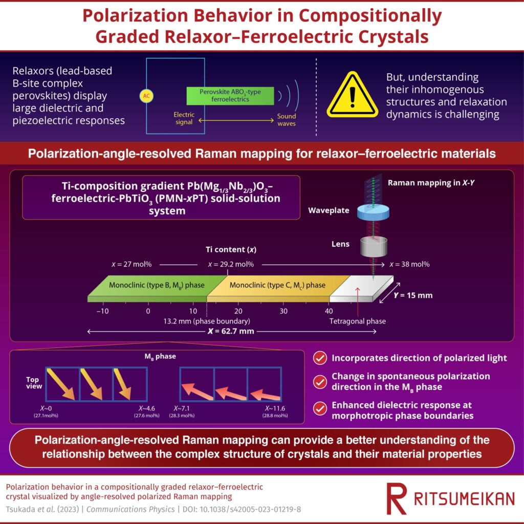
One characteristic feature of PMN-PT crystals is their dielectric and piezoelectric response at the boundaries that separate the different phases of the material. The specific structure of the PMN-PT crystal, especially the amount of titanium (Ti), can affect the structure and characteristics of the phase boundaries. To investigate the effect of Ti mixing ratios on dielectric materials, the researchers photographed a crystal sample of 62.7 × 15.0 × 0.3 PMT-PT with the newly installed Raman map in the microscope.
The Ti content varies from 27.0 mol% to 38.0 mol% along the length of the sample, which resulted in three distinct phases: the monoclinic phase (type B) where the Ti content was 27 mol% and 29.2 mol%, The monoclinic (type C) phase increased to 34.5 mol%, and the tetragonal phase with a Ti content of 34.8-38.0 mol%.
By examining the Raman spectra corresponding to the different light spectra of each part of the sample, the researchers observed a sudden change in the intensity of the Raman peaks only in the B phase of type B. In addition, they also observed a different change following the spontaneous polarization in this phase. The spectra revealed a gradual relaxation (reorientation of electric dipoles in response to thermal disturbances) of the polarization of the material near the phase boundary between the monoclinic phases (type B) and (type C). This, in turn, indicates that the reorientation of the dipoles occurs at a lower rate, resulting in the material retaining more charge and exhibiting a stronger dielectric response at the phase boundary.
“We found that the ability of the relaxor-ferroelectric material to store large amounts of electricity due to the slow response of the nanometer-scale electric polarization to the external voltage,” explains Prof. Fujii.
In summary, the analysis of the optical properties of relaxation devices shows the potential of dynamic polarization-angle-resolved polarization microscopy, which can help improve dielectric performance. In particular, the understanding of the polarization behavior of PMT-PT can improve the development of regenerative devices with better ultrasound detection and next-generation imaging systems.
More information:
Shinya Tsukada et al, Polarization behavior in a stable relaxation-ferroelectric crystal observed by static Raman mapping, Communications Physics (2023). DOI: 10.1038/s42005-023-01219-8
More news:
Communications Physics
Provided by Ritsumeikan University
#spectroscopic #method #investigation #relaxorferroelectric #materials