Molecular scientists have long believed that euchromatin, the part of chromatin that is made up of genes and is active for genes, is open and can be transcribed. A research team, looking at new evidence from genomics and comparative studies, has written a conceptual paper showing that euchromatin is not open. The paper is published in Trends in Cell Biology.
In their paper the group discusses their new concept of euchromatin in the cell and shows how the revealed organization works on the genome. “Our main goal is to reveal how genomic information is analyzed and read in living cells,” said Kazuhiro Maeshima, lead author and professor at the National Institute of Genetics and SOKENDAI, Japan.
Chromatin describes the combination of DNA and proteins in human cells and other eukaryotes. According to examples in the literature, chromatin exists in two forms—euchromatin, which is very compact and can be transcribed, and heterochromatin, which is compact and usually not transcribed. Transcription is the process by which a cell makes an RNA copy of a DNA molecule, an essential process for life. The research team focuses their research on euchromatin.
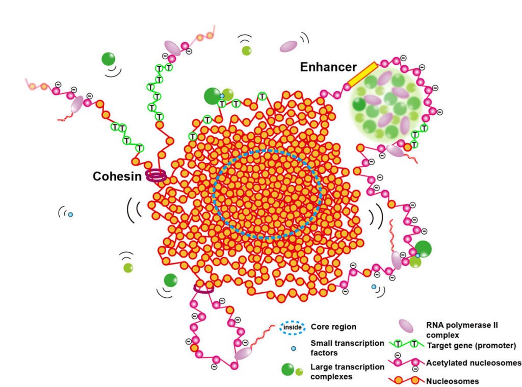
The group shows in their paper that euchromatin in higher eukaryotic cells like human cells is not open. Instead, euchromatin is thought to form fluid-like spheres, with sizes ranging from 100 to 300 nm in diameter.
Recent studies involving 3D simulated illumination microscopy (SIM), ion beam scanning electron microscopy, and single-molecule imaging have revealed local chromatin conformation and have shown evidence that euchromatin forms compact regions. In addition to this, recent genomics techniques have also revealed that only a few regions are always present.
The research shows that the condensed domains are unstable and strongly bend and have fluid-like properties. Euchromatic regions are smaller in size than heterochromatic regions, resulting in a higher phase-to-volume-ration in euchromatin, which may also explain differences in transcriptional activity.
“The open areas of chromatin where transcription takes place appear to be limited to domains or boundaries between domains. Also, the condensed domains have fluid-like properties, which provide access to the interior, and allow cells to initiate transcription and other DNA events, such as replication of DNA is repair,” said Shiori Iida, co-first author.
Chromatin cannot be divided into euchromatin and heterochromatin with its open/closed structure. Contrary to the accepted theory, euchromatin regions are not open, but shortened. “Condensed chromatin appears to be a stable chromatin state in higher eukaryotic cells, possibly active units in DNA transactions, and compact units like Lego blocks of chromosomes during cell division,” said Kazuhiro Maeshima.
Looking to the future, the team hopes that their work will expand the understanding of the coding and other processes of DNA. “The next step is to investigate how the shortened regions are regulated during cell differentiation or developmental processes to perform certain cellular functions,” said Masa Shimazoe, one of the co-authors.
More information:
Trends in Cell Biology (2023). DOI: 10.1016/j.tcb.2023.05.007
Presented by the Research Organization of Information and Systems
#group #presents #perspective #euchromatin #cell


